 When your doctor orders an imaging test, you may well wonder why they select one type of imaging over the other. MRI and CT (or CAT) scans can be used for similar purposes, and several factors play into the decision to use one over the other.
What is a CT scan?
A CT scanner uses X-rays to generate detailed images for close examination. X-ray beams and detectors rotate around the patient to create cross-sectional images of the body that can be reformatted to produce three-dimensional visualizations of the features being examined.
How are CT scans used?
CT scanning is ideal for getting detailed images of bony structures as well as imaging bone, soft tissue, and blood vessels all at once. CT is capable of producing good soft tissue differentiation, as well, particularly when IV contrast is used. CT is commonly used for diagnosing lung and chest problems and detecting cancers, and emergency rooms rely heavily on CT scanning because it produces fast, detailed results.
What are the drawbacks of CT?
CT offers less precise visualization of soft tissue detail and subtle differences in soft tissues than MRI. However, CT is still often the preferred choice for some soft tissue examinations because of other benefits, such as speed, cost, and availability.
CT uses ionizing radiation, which is associated with an increased risk of cancer. However, depending on the type of CT examination performed, the radiation dose involved often results in only a very low increase in lifetime fatal cancer risk. For a better understanding of the radiation dosages involved with various CT procedures and their associated risks, see our article, “Wise and Safe Diagnostic Imaging: Understanding Radiation Dose.” Any time your doctor orders a procedure that uses ionizing radiation, it should be because the benefit of the results obtainable from the exam outweighs the risk the exam poses. If you ever have questions about this risk/benefit balance, ask!
Finally, the contrast solution that is used with CT typically contains iodine, which can cause allergic reactions in some patients. While such reactions are rare, they are less rare in response to iodine-containing contrast solutions than to other types of contrast used with MRI.
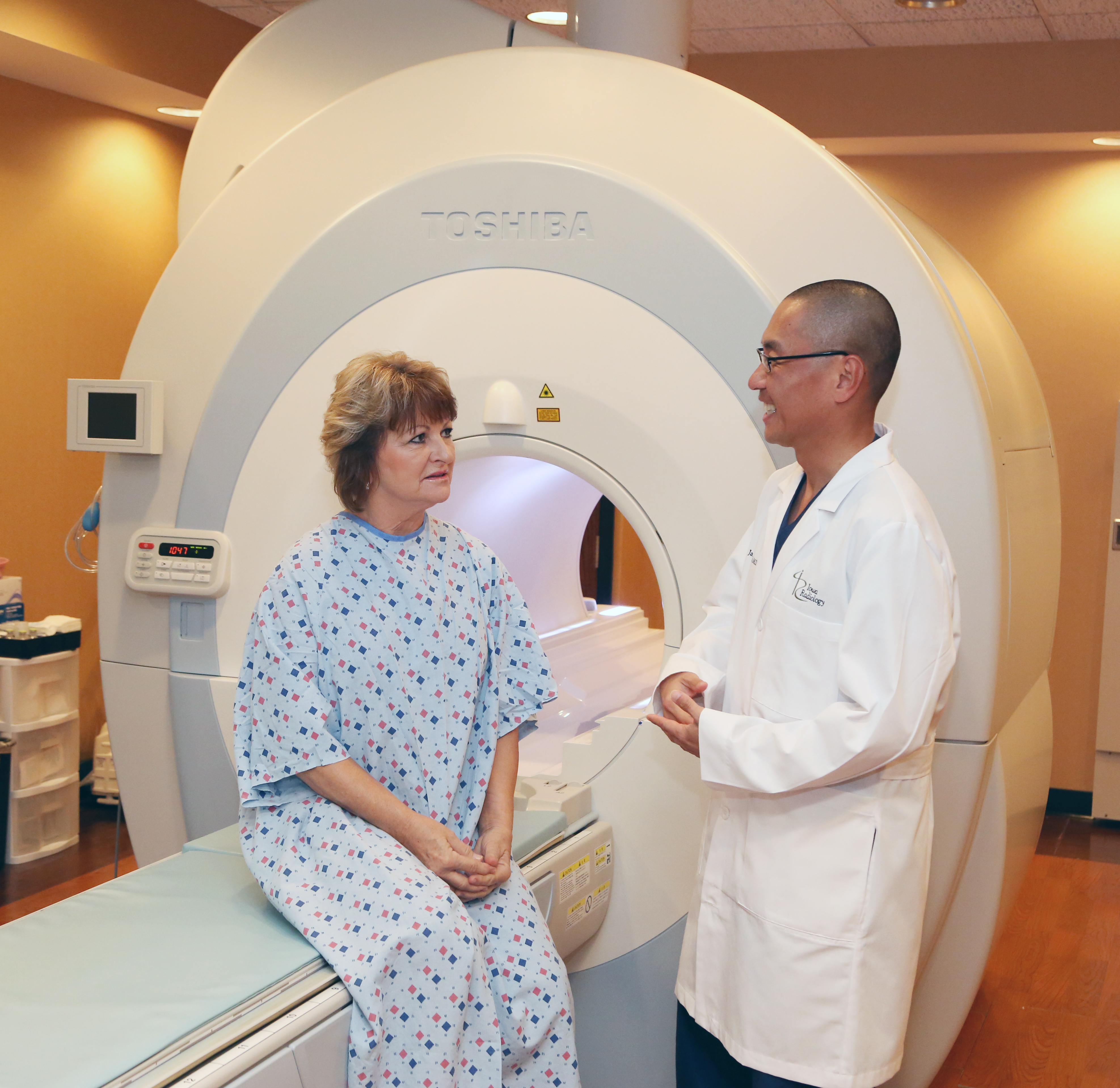
What is MRI?
Magnetic resonance imaging uses a powerful magnetic field to produce detailed pictures of the body’s interior structures. The patient lies on the exam table, which slides into a tube in the center of the magnet. Different types of MRI machines are available, with different types of bores—traditional, short, open, or wide—to accommodate different patient needs. For example, a wide-bore MRI will be more comfortable for claustrophobic or larger-bodied patients than a traditional bore.
{{cta(‘e90cab36-5b00-4024-ae3c-a3c900ed4571’)}}
How is MRI used?
MRI is the star when it comes to close examination of soft tissue detail. MRI technology allows radiologists to adjust the contrast in images, enabling them to more clearly visualize the differences between various types of soft tissue. As a result, MRI is a procedure of choice for examining ligament and tendon injuries, spinal cord injuries, and brain tumors, and it can produce superior images of other types of tumors as well. Because MRI doesn’t use ionizing radiation, it can provide the benefit of detailed imaging without increasing a patient’s cancer risk.
What are the drawbacks of MRI?
MRI provides less detail of bone than CT does, and images are less detailed overall because they are taken over a much longer time span. While a CT scan often takes less than five minutes from start to finish, MRI requires the patient to remain motionless for prolonged periods in order to obtain usable images. A typical MRI exam takes 30–60 minutes, but some can take up to two hours in total. Not only does this result in blurring due to unavoidable small movements, but it also makes MRI unsuitable for time-critical emergency evaluations and can cause anxious or claustrophobic patients to find the procedure difficult to endure. When necessary, doctors can prescribe sedatives to enable these patients to complete an MRI exam.
Additionally, some patients are not appropriate candidates for MRI. The powerful magnet used makes MRI unsuitable for patients with implanted ferromagnetic elements such as aneurism clips, pacemakers, cochlear or ocular implants, and imbedded shrapnel. Because the magnetic field is incredibly strong, it is vitally important that you understand all warnings that your clinic provides prior to the exam about what can and cannot be brought into the exam room. Learn more about contraindications to MRI in our previous article.
Finally, MRI is more costly and less widely available than CT. Because CT provides many of similar benefits to MRI at around half the cost, it is often a more practical choice for patients’ imaging needs.
{{cta(‘4122f3d3-66b7-4250-98c7-edb67ef514fa’,’justifycenter’)}}
Iowa Radiology provides a wide variety of imaging services, including CT, MRI, ultrasound, low-dose 3-D mammography, and much more. For more information about MRI and CT exams, click below to access our free eBooks.
{{cta(’38e891b6-c06e-40d4-88c2-104d4672ad39′,’justifycenter’)}}
The information contained in the Iowa Radiology website is presented as public service information only. It is not intended to be nor is it a substitute for professional medical advice. You should always seek the advice of your physician or other qualified healthcare provider if you think you may have a medical problem before starting any new treatment, or if you have any questions regarding your medical condition.
Iowa Radiology occasionally supplies links to other web sites as a service to its readers and is not in any way responsible for information provided by other organizations.
Recent Comments