

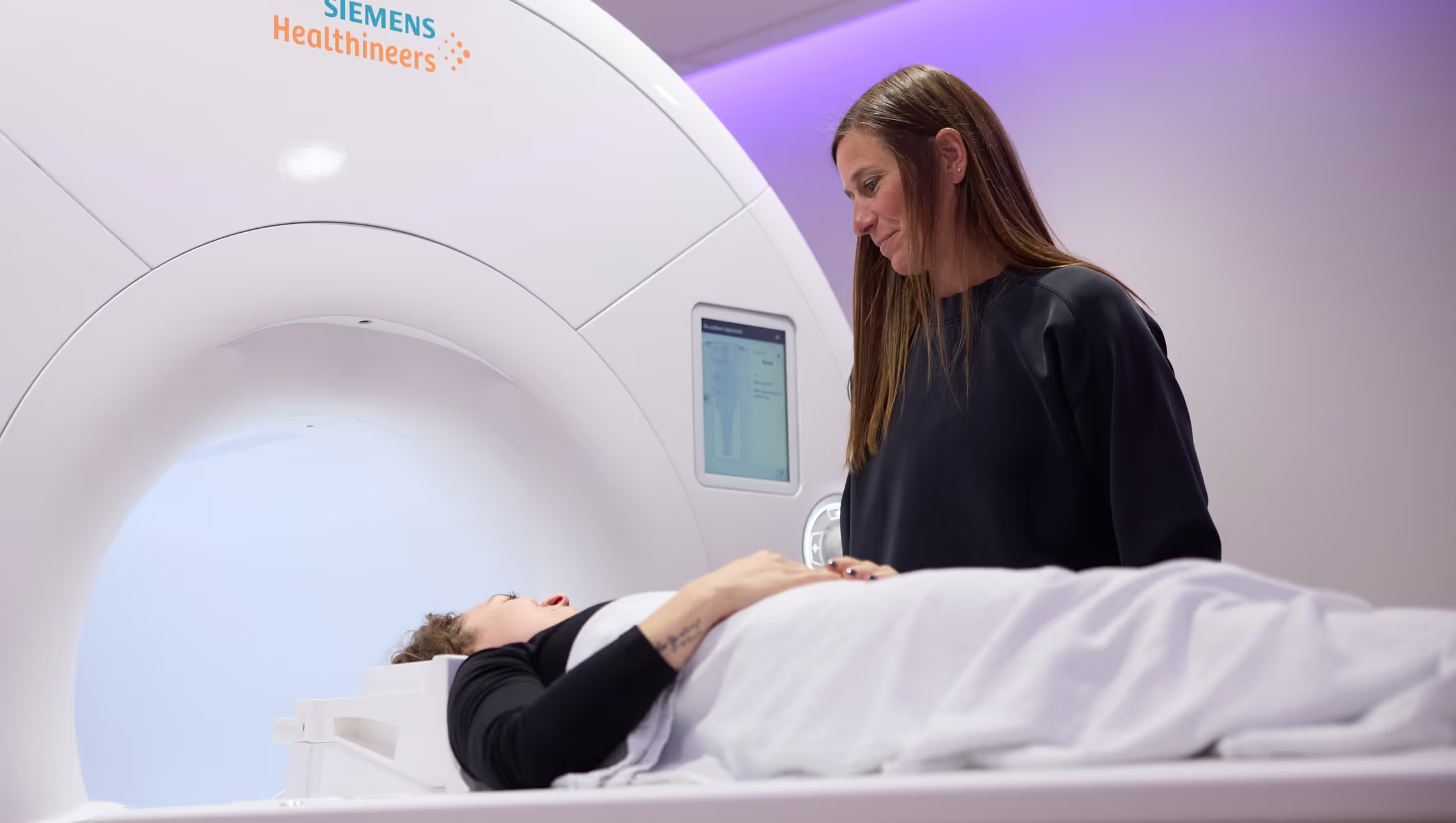
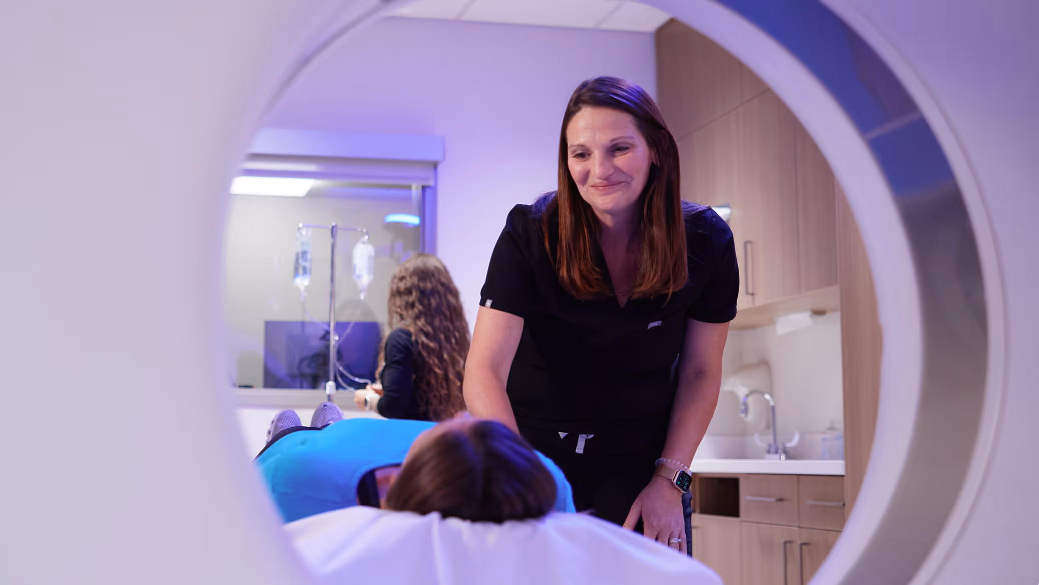
What is an Abbreviated Breast MRI?
MR imaging uses a magnetic field, radio waves, and a computer to produce detailed images of your breast. MRI does not use radiation. Your doctor will likely order this exam for implant integrity evaluation, assessment of newly diagnosed breast cancer or post lumpectomy, specialized follow up, or evaluation of very high-risk patients whose lifetime risk of breast cancer is greater than 20%. Calculate your lifetime risk here. High risk breast MRI is generally covered by insurance. Because your comfort is important to us, we provide music headsets as well as blankets to make your experience more comfortable. Our knowledgeable, professional team will put you at ease and answer all of your questions before the exam.
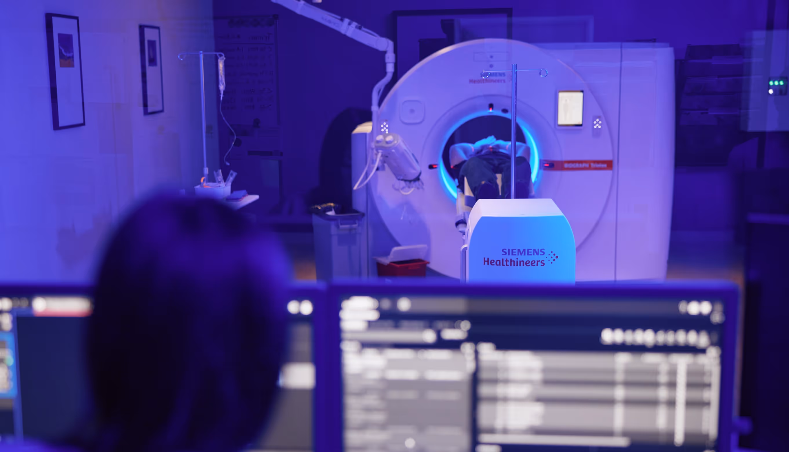
What happens during the test?
Our technologist will take a brief medical history. You will be asked to lie down on your stomach on an exam table, where your breasts will hang into cushioned openings. The table will then slide into the scanning area. During the test, the MRI will make a rapid tapping noise. Just relax and remain still. Initial images will be obtained, and then contrast material will be injected into a vein in your arm. After injection, additional images will be taken.
You should plan 45 minutes of total clinic time. The scan typically takes 10-15 minutes. You may return to normal activities following your exam.
How do I prepare for the test?
Typically, no preparation is required before the exam. No fasting is necessary. For premenopausal women, this exam is scheduled 6-10 days after the first day of your period. You will need to remove all jewelry, hairclips, and bobby pins. In addition, you will need to remove your bra and any clothing containing metal. You will be provided a gown and a secure locker in which you can place valuables. You may take oral anti-anxiety medication as prescribed by your doctor. Iowa Radiology does not administer or prescribe anti-anxiety medications. Please ensure you have a driver.
When can I expect the results?
A radiologist will review the images and send a report to your referring physician within two business days. Your doctor will review the report and contact you with the results.
What contraindications should I be aware of?
If you are pregnant, have had an aneurysm clip, have had ear or eye prosthesis, or have a pacemaker, you may not be a candidate for an MRI.
Downloadable Resources
FAQ's
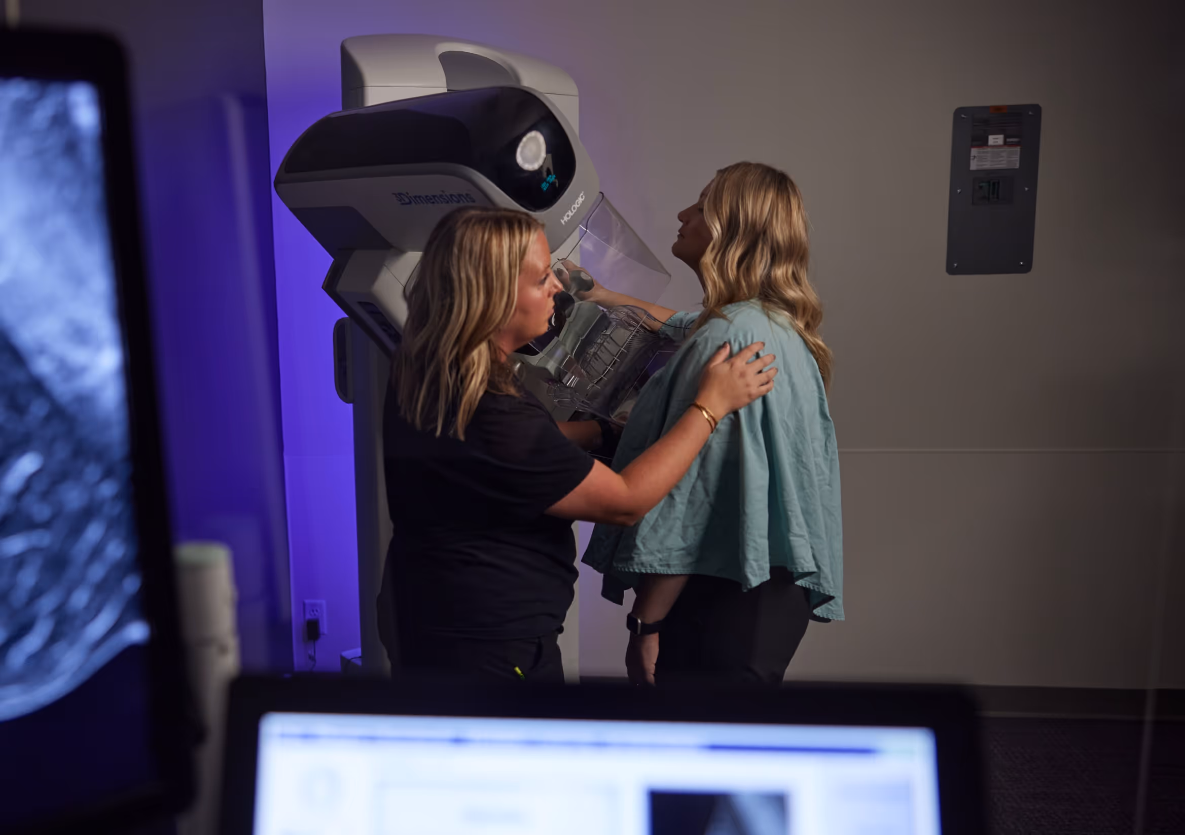
What is breast density, classification, and significance?
Breasts are made up of fibrous, glandular and fatty tissue. Your breasts are considered dense if you have a lot of fibrous or glandular tissue and not much fat. Breast density is determined by the radiologist when reading your mammogram. Approximately 50% of women have dense breasts (C or D). Dense breasts may make cancer detection more difficult.
Density is determined using a 4- level scale:
A- Almost entirely fatty – 10% of women are in class A
B- Scattered areas of fibro glandular density – 40% of women are in class B
C- Heterogeneously dense – 40% of women are in class C
D- Extremely Dense – 10% of women are in class D
What should I do if I have dense breasts?
Beginning at age 40, get a yearly mammogram. A mammogram is the only medical imaging screening test proven to reduce breast cancer deaths. It is the gold standard screening tool and many cancers are found because of this exam. Iowa Radiology has adopted 3D digital mammography as standard of care because it has been proven to reduce callbacks and to detect breast cancers much earlier than traditional 2D mammograms.
If you do or don’t have dense breasts, talk to your doctor. Breast density is just one determining factor that may place you at increased risk of developing breast cancer. You and your doctor can decide if additional testing is appropriate for you.
How has Iowa law changed breast density reporting?
Iowa Radiology has always reported breast density to your referring provider and beginning in January 2018, in accordance with the changes in Iowa law, we also began reporting density to you in your results letter.
What are the risk factors for breast cancer?
- Overweight after menopause
- Advancing Age
- Diet
- Sedentary lifestyle
- Breast cellular changes
- Nulliparity
- Decreased vitamin D levels
- Exposure to chemicals
- Race/ Ethnicity
- Alcohol Consumption
- Being a woman
- Smoking
- Having dense breasts
- Genetic mutation
- Taking hormones
- Family history of ovarian cancer
- Family history of breast cancer
- Personal history of breast cancer
- Advanced maternal age > 30
- Never breast feeding
- Genetic mutation
- Menstruation < 12 years of age Menopause > 55 years of age
- Lifetime breast cancer risk > 20%
- Certain benign breast changes
- Chest or face radiation after age 30
- DES drug exposure (diethylstilbestrol)
Iowa Radiology routinely calculates your lifetime risk of breast cancer which is included in our report to your provider.
How often is breast MRI recommended?
The American College of Radiology (ACR) recommends that high risk breast MRI’s be performed every 12 months. Patients at average risk (below 20% lifetime risk) may benefit from abbreviated breast MRI (AB-MRI) every 1-3 years to supplement a yearly screening mammogram.
Where we offer
Breast MRI
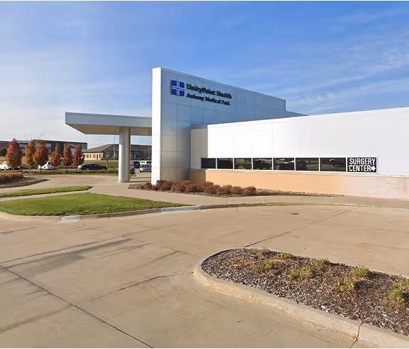
3625 N Ankeny Blvd. Suite H Ankeny, IA 50023
Mon – Fri: 8am-5pm
Sat: Hours available for MRI and Digital Mammography
Fax: (515) 963-7619

12368 Stratford Drive Suite 300 Clive, IA 50325
Mon – Fri: 6:30am–5pm
Sat: Hours available for MRI & Digital Mammography
Fax: (515) 226-8408

2515 Grand Prairie Parkway, Waukee, IA 50263
Mon – Fri: 8am-5pm
Fax: (515) 226-8408
Featured Articles


Schedule an Appointment
At Iowa Radiology, we strive for excellence in everything we do. You can expect easy access, convenient scheduling, and timely service.
Schedule an Appointment
At Iowa Radiology, we strive for excellence in everything we do. You can expect easy access, convenient scheduling, and timely service.