Advances in breast imaging continue to improve how radiologists detect and evaluate breast abnormalities, giving patients and providers more tools to support informed care decisions. One of the newer imaging options now available at Iowa Radiology is Contrast-Enhanced Mammography (CEM), an advanced imaging exam used in select situations when additional breast evaluation is needed.
While breast MRI remains the most established and evidence-based option for supplemental screening in women at high risk for breast cancer, CEM can provide an important alternative for patients who may not be candidates for MRI.
What Is Contrast-Enhanced Mammography?
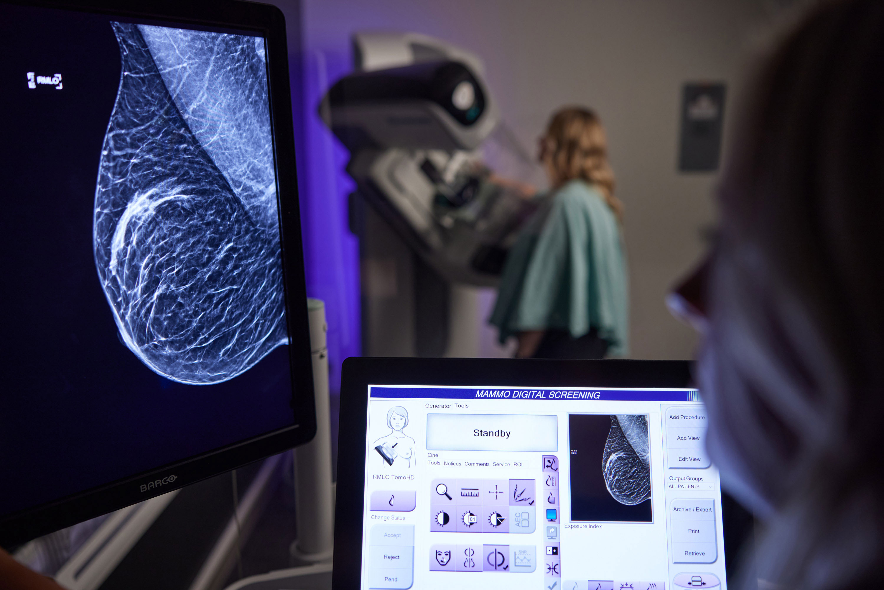
Contrast-Enhanced Mammography combines traditional mammography with an intravenous contrast agent to help highlight areas of increased blood flow within the breast. Because cancers often develop increased blood supply as they grow, contrast-enhanced imaging can help radiologists identify areas that may require closer evaluation.
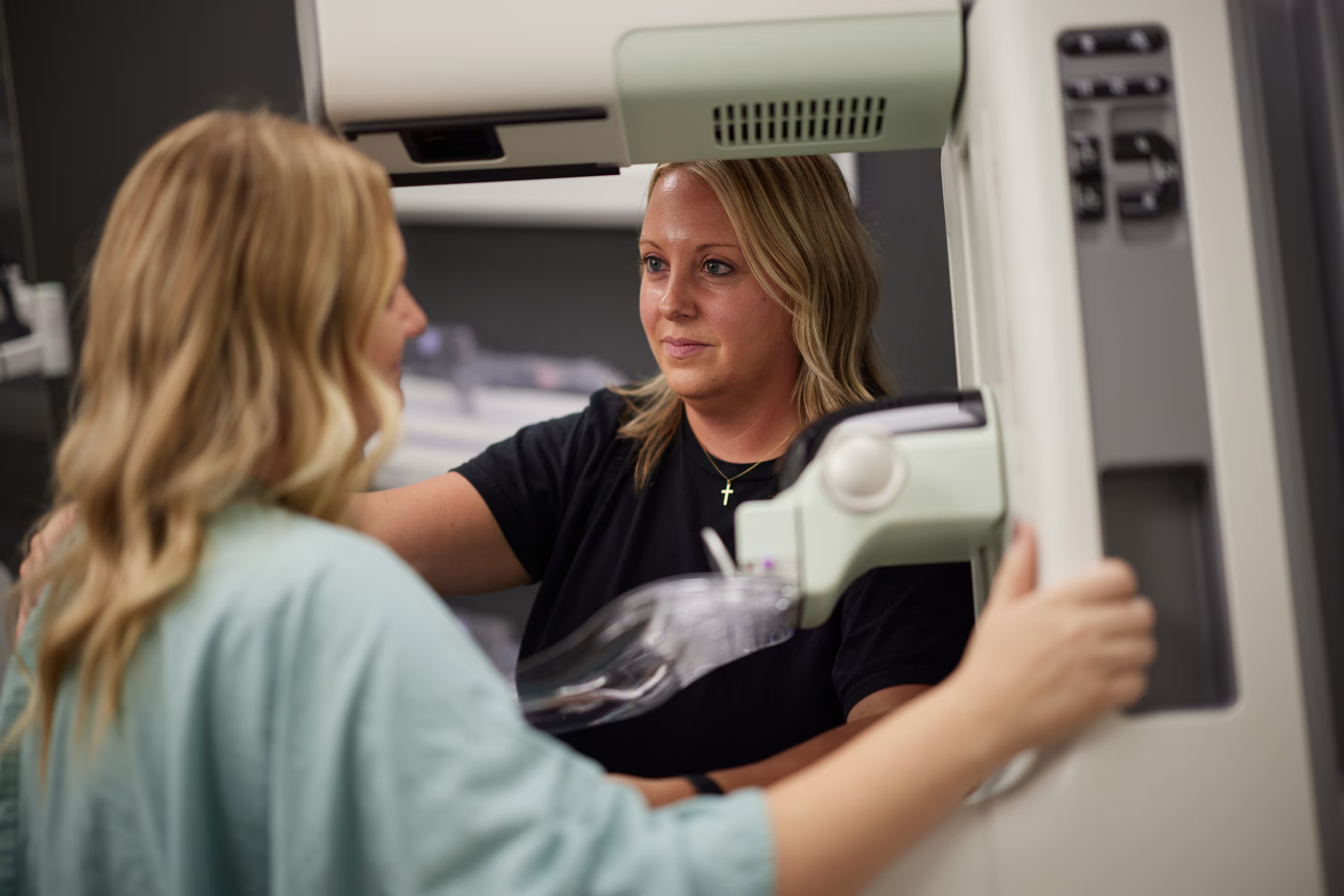
During the exam, contrast material is injected into a vein before specialized mammogram images are obtained. The exam itself is performed similarly to a standard mammogram, though the addition of contrast provides radiologists with more detailed diagnostic information.
When Is CEM Used?
CEM is not intended to replace breast MRI for high-risk breast cancer screening. According to current breast imaging recommendations, breast MRI remains the preferred supplemental screening tool for women with a lifetime breast cancer risk greater than 20%, often alternating with screening mammography every six months.
However, CEM may be considered in select situations, including:
- Patients who cannot safely undergo MRI because of certain implanted medical devices or other contraindications
- Patients who experience significant claustrophobia during MRI exams
- Situations where additional diagnostic clarification is needed and MRI is not feasible
- Certain high-risk patients who need an alternative imaging option
CEM is not recommended as routine supplemental screening for women at average risk for breast cancer.
How Does CEM Differ From Breast MRI?
Both breast MRI and CEM use contrast material to evaluate breast tissue in greater detail than standard mammography alone. Breast MRI uses magnetic fields and radio waves to create highly detailed images of the breast without radiation exposure.
CEM, by comparison, combines mammography technology with contrast enhancement to provide additional imaging detail when needed. While MRI remains the most comprehensive and well-studied supplemental imaging option for high-risk patients, CEM serves as a valuable alternative in carefully selected cases.
The most appropriate exam depends on several factors, including a patient’s medical history, breast cancer risk level, prior imaging results, and ability to safely undergo MRI.
Delivering Insights That Guide Care
At Iowa Radiology, experienced breast imaging specialists work closely with referring providers to help ensure each patient receives the most appropriate imaging for their individual needs. Our women’s imaging services combine advanced technology, expert interpretation, and a patient-centered approach focused on clear communication and compassionate care.
Iowa Radiology offers Breast MRI at multiple locations throughout the Des Moines area, along with Contrast-Enhanced Mammography (CEM) at our Clive location. Patients should speak with their provider about which imaging option may be most appropriate for them.